Digital Imaging Technology: the latest in eye care advancements
 The eye doctors at Armstrong & Small Eyecare Centre use cutting-edge digital imaging technology to assess your eyes. Many eye emergencies and eye diseases, if detected at an early stage, can be treated successfully without a loss of vision. Your retinal Images will be stored securely electronically. This gives the eye doctor a permanent record of the condition and state of your retina or what we like to refer to as a “digital library” of your eye health.
This is very important in assisting your optometrist in Winnipeg to detect and measure any changes to your retina each time you get your eyes examined, as many eye conditions, such as glaucoma, diabetic retinopathy and macular degeneration are diagnosed by detecting changes over time.
The advantages of digital imaging include:
- Quick, safe, non-invasive and painless
- Provides detailed images of your retina and sub-surface of your eyes
- Provides instant, direct imaging of the form and structure of eye tissue
- Image resolution is extremely high quality
- Uses eye-safe near-infra-red light
- No patient prep required
Optos Ultra-Wide Retinal Exam in Winnipeg, MB
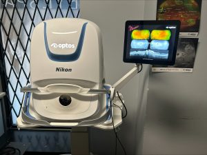 At Armstrong & Small Eyecare Centre, we are proud to be one of the first eyecare clinics in Manitoba to use the Optos Ultrawide Image Scanner starting back in 2004. The Monaco is our latest Ultra-Wide Retinal Imaging device that allows our eye doctor to evaluate the health of the back of your eye, the retina. It is critical to confirm the health of the retina, optic nerve and other retinal structures. The digital camera snaps a high-resolution, ultra-wide digital picture of your retina. This picture clearly shows the health of your eyes and is used as a baseline to track any changes in your eyes in future eye examinations at our optometry practice in Winnipeg.
The Monaco not only offers coloured scans of your retina, but also fundus auto-fluorescent (FAF) images that help your optometrist in Winnipeg to detect retinal diseases at their earliest stages. The Monaco also features a built-in OCT (Optical Coherence Tomography) that allows your optometrist in Winnipeg to have even more detail of the delicate structure of your retina.
All of our adult eye exams include the Optomap® Retinal Exam as an important part of our comprehensive eye exams. The Optomap® Retinal Exam produces an image that is as unique as your fingerprint and provides us with a wide view to look at the health of your retina. The retina is the part of your eye that captures the image of what you are looking at, like film in a camera. As this technology was created to be able to better image the retinas of children, we also recommend imaging for anyone under the age of 18 every 3 years.
Many eye problems can develop without you knowing. You may not even notice any change in your sight. But diseases such as macular degeneration, glaucoma, retinal tears or detachments, and other health problems such as diabetes and high blood pressure can be seen with a thorough exam of the retina.
An Optomap® Retinal Exam provides:
- A scan to show a healthy eye or detect disease.
- A view of the retina, giving your doctor a more detailed view than he/she can get by other means.
- The opportunity for you to view and discuss the Optomap® image of your eye with your doctor at the time of your exam.
- A permanent record for your file, which allows us to view your digital personalized library each year to look for changes
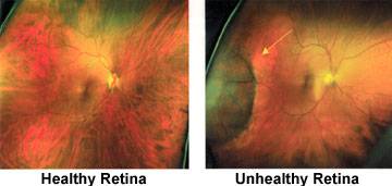
The Optomap® Retinal Exam is fast, easy, and comfortable for all ages. To have the exam, you simply look into the device one eye at a time and you will see a comfortable flash of light to let you know the image of your retina has been taken. The Optomap® image is shown immediately on a computer screen so we can review it with you.
Please schedule your Optomap® Retinal Exam today!
Optical Coherence Tomography (OCT)
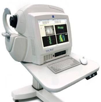 An Optical Coherence Tomography scan (commonly referred to as an OCT scan) is the latest advancement in imaging technology. Similar to an ultrasound, this diagnostic technique employs light rather than sound waves to achieve higher resolution pictures of the structural layers of the back of the eye.
We are proud to have the latest and most advanced in OCT technology - the Zeiss Cirrus HD OCT 5000.
A scanning laser used to analyze the layers of the retina and optic nerve for any signs of eye disease, similar to a CT scan of the eye. It works using the light without radiation and is essential for early diagnosis of glaucoma, macular degeneration, and diabetic retinal disease.
With an OCT scan, doctors are provided with color-coded, cross-sectional images of the retina. These detailed images are revolutionizing early detection and treatment of eye conditions such as wet and dry age-related macular degeneration, glaucoma, retinal detachment, and diabetic retinopathy.
An OCT scan is a non-invasive, painless test. It is performed in about 10 minutes right in our office. Feel free to contact our office to inquire about an OCT at your next appointment.
Visual Field Testing
 A visual field test measures the range of your peripheral or “side” vision to assess whether you have any blind spots (scotomas), peripheral vision loss or visual field abnormalities. It is a straightforward and painless test that does not involve eye drops but does involve the patient's ability to understand and follow instructions. This instrument is used most often for patients who have, or may be suspicious of, glaucoma. It is also used for patients who may be dealing a neurological change such as a stroke, those who are taking Hydroxychloroquine (Plaquenil) or to monitor changes that may affect your driving capabilities.
Our special equipment might be used to test your visual field. In one such test, you place your chin on a chinrest and look straight ahead at a fixation target. Lights are flashed on, and you must press a button whenever you see the light. The lights are bright or dim at different stages of the test. Some of the flashes are purely to check you are concentrating. Each eye is tested separately, and the entire test takes 10-20 minutes. These machines can create a computerized map out your visual field to identify if and where you have any deficiencies.
Nidek OPD Scan
 |
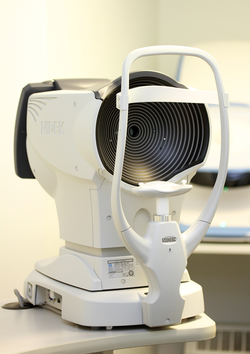 |
The Nidek OPD III refractive power and the corneal analyzer is the latest instrument acquired by Armstrong & Small Eyecare Centre to provide our doctors with the best information about your prescription and cornea. The Nidek OPD III acts as both a wavefront aberrometer and a corneal topographer.
Wavefront Aberrometer
Wavefront aberrometry gives an unprecedented assessment of visual acuity and quality of vision. Our machine can detect higher order aberrations in the optical pathway between the cornea and the retina. This allows for precise quantification of refractive error and aids in determination of optimal corrective eyewear.
Topographer
Corneal topography provides intuitive elevation maps and numerical data for the corneal surface. This allows for enhanced detection of corneal pathology such as keratoconus and pellucid marginal degeneration. Serial topography is key in determining if such conditions are progressing over time which may indicate the need for surgical intervention.
iCare Tonometer
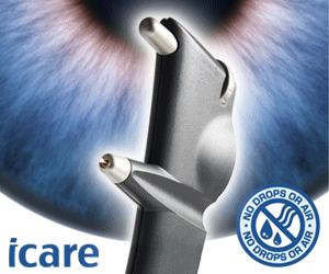 iCare Tonometers are used for easy, accurate and patient-friendly intra-ocular pressure measurement, which is used for glaucoma screening. iCare tonometers are based on unique, patented rebound technology, in which a very light and small probe is used to make a momentary contact with the cornea. The iCare tonometer is based on a proven accurate measuring principle, in which a very light probe is used to make momentary and gentle contact with the cornea. The measurement is barely noticed by the patient. The device not only makes IOP measuring a more pleasant experience on all patients, it is also an important break-through for succeeding with non-compliant patients (i.e. children and dementia patients).
Mediworks Firefly Digital Slit Lamp and Meibographer
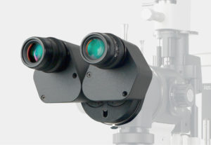 Every optometrist uses a slit lamp to assess the front of the eye during an eye exam, but with our new “Firefly” slit lamp also has built-in technology to assess dry eye disease. We are now able to not only capture high-resolution images of such structures as the eyelids, cornea (clear front cap), iris (colored part of your eye) and crystalline lens, but we can also assess the structure of your meibomian glands within the eyelid itself. Just like the Optomap being able to image your retinas in detail, being able to show you instant images of our concerns relating to the front of the eye, is also a crucial step in our high-level eye care assessment and your own education.
The FireFly has a built-in infra-red imaging system that allows your optometrist to image the structures within the lower and upper lids known as meibomian glands. The meibomian glands are essential oil glands that function by producing crucial oil to the tears that help the tear film to evaporate more slowly from the surface of the eye. Being able to image these structures during a dry eye assessment is a crucial step in determining the type of dry eye that you may be dealing with and will help your optometrist determine the best treatment strategy for you.
The FireFly slit lamp not only contains a meibomian gland observation, but also software that will further assess your potential dry eye with such features as non-invasive tear breakup time (how quickly are your tears evaporating off the surface of your eye), tear meniscus height (how your tear film is positioned on your lower lid) and red eye analysis. Each of these components will guide your optometrist in evaluating the best next steps in treating your dry eye disease.
